Spine Fractures
What are common spine fractures?
Two common fractures are a result of
- Trauma (fall from height or high speed motor vehicle accident)
- Osteoporesis
What are Vertebral Compression Fractures?
Vertebral compression fractures occur when the normal vertebral body of the spine is squeezed or compressed. In younger people, the bone can resist the force, but as we get older, the bone becomes more brittle and can break with normal forces. The bone collapses when too much pressure is placed on the vertebrae, resulting in pain, limited mobility, loss of height and spinal deformity. In very severe compression fractures, the back of the vertebral body is pushed into the spinal canal and pressure is placed on the spinal cord.
Causes of Vertebral Compression Fractures
Vertebral compression fractures can occur because of trauma but the most common cause is due to aging. Aging results in osteoporosis, a condition that causes thinning of the bone. The thinning bones can cause tiny fractures during normal activities.
Symptoms of Vertebral Compression Fractures
The most common symptom of a vertebral compression fracture is severe pain in your back, which worsens on standing or walking and decreases when resting. You may also feel weakness and numbness in the affected areas, disability, and limited spinal mobility.
Diagnosis of Vertebral Compression Fractures
Your doctor will carefully examine you based on the symptoms and medical history. Your doctor may also recommend other diagnostic tests such as:
- X-ray: A spinal X-ray may be ordered to determine the presence of a fracture.
- MRI scan: An MRI of the spine may be performed to determine if the fracture is old or new, and to detect other soft tissue abnormalities.
- Bone scan: A nuclear bone scan may be ordered to help determine the presence or age of the fracture.
- DEXA scan: Dual-energy X-ray absorptiometry (DEXA) scan is a test to measure bone mineral density, and is typically ordered to diagnose osteoporosis.
Treatment of Vertebral Compression Fractures
The treatment of vertebral compression fractures aims at reducing the pain and stabilizing and repairing the fracture. This is best achieved by medications, back braces, bed rest and physical therapy.
Your doctor may prescribe a back brace that supports the back and restricts movement.
Surgery may be suggested if you continue to have severe pain despite non-surgical treatment. Two minimally invasive surgical procedures for treating vertebral compression fractures include:
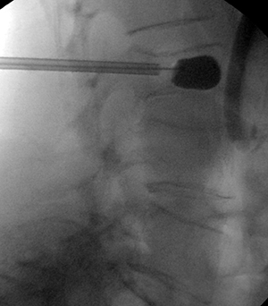
Kyphoplasty: In this procedure, a large needle is introduced into the spine with a balloon called a bone tamp and inflated until it expands to the desired height. The space created is then filled with or injected with orthopedic cement called polymethylmethacrylate (PMMA). This procedure restores the height of the vertebrae and reduces the deformity.
Vertebroplasty: This technique involves inserting polymethylmethacrylate into the bone of the collapsed vertebra with the needle and syringe under the guidance of X-ray. This technique is performed to stabilize the fracture and prevent further collapse.
Prevention of Vertebral Compression Fractures
The most effective way to prevent vertebral compression fractures is to prevent osteoporosis. A well-balanced diet, regular exercise program, calcium and vitamin D supplements, smoking cessation, estrogen hormone replacement for women and practicing good posture may help you to prevent osteoporosis. The most effective way, however, is to be placed on osteoporosis medications. Without medication, your risk for another fracture within a year may be as high as 30%.
Clinical Example 1
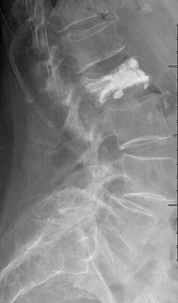
88 year old female fell in the bathroom with increasing amount of back pain. Despite pain pills in the hospital, she could not get out of bed. MRI showed compression fracture due to osteoporosis (weak bone). MRI can show if the fracture is new based on swelling in the bone. The arrow shows the bone is whiter than the other bones indicating a new fracture.

She underwent minimally invasive kyphoplasty to treat the fracture. Through a pen hole incision a balloon is placed inside the bone to restore the bone back to normal shape as possible. (It never looks quite normal). Then cement is placed into the bone and acts like grout making the fracture all healed.
Clinical Example 2
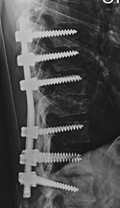
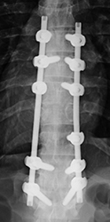
Gentleman after a motor vehicle accident comes into the hospital unable to move his legs. A CT shows a fracture dislocation (arrow). Emergent surgery is done to help realign the fracture as best as possible.

You will need the Adobe Reader to view and print these documents.![]()