Kyphoplasty
Balloon Kyphoplasty
What are Vertebral Compression Fractures
Osteoporosis (bone disease) is the primary cause of vertebral compression fractures. Other causes include trauma such as a fall or motor vehicle accident and some types of cancers affecting the spinal vertebrae.
What is Balloon Kyphoplasty?
Balloon kyphoplasty is a spine surgery that relieves back pain caused by a vertebral compression fracture. The aim of balloon kyphoplasty is to relieve pain, stabilize the fracture and restore the vertebral body height.
Indications of Balloon Kyphoplasty
Your doctor recommends balloon kyphoplasty if you have severe pain and deformity that is not relieved by non-surgical treatment modes including rest, pain medications and braces.
Balloon Kyphoplasty Procedure
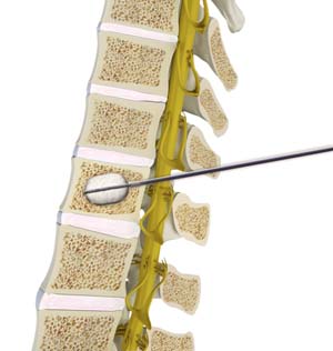
The surgery is performed under sedation/twilight sleep. During the procedure, you will lie face down on the operating table. A small incision is made in the back through which a narrow tube is inserted into the compressed vertebra under the guidance of live X-ray. Then a special balloon is inserted through this tube and carefully inflated. This elevates the fracture and restores the vertebra to its original height. The balloon is then deflated and removed, leaving behind an open cavity. The cavity is filled with bone cement with the help of miniature surgical instruments. The cement hardens within a few minutes and stabilizes the bone.
What to Expect after Balloon Kyphoplasty
You may experience significant pain relief following surgery and will be allowed to get up and walk. Your doctor will prescribe pain medication if necessary and recommend a rehabilitation program to strengthen your spinal muscles. You should avoid strenuous activities for at least 6 weeks.
Risks and Complications of Balloon Kyphoplasty
As with any surgery, balloon kyphoplasty may be associated with certain complications such as infection, nerve or spinal cord injury and cement particles entering the blood or spinal fluid. There is also a risk of leakage of cement outside of the bone causing nerve damage.
Clinical Example
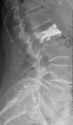
88 year old female fell in the bathroom with increasing amount of back pain. Despite pain pills in the hospital, she could not get out of bed. MRI showed compression fracture due to osteoporosis (weak bone). MRI can show if the fracture is new based on swelling in the bone. The arrow shows the bone is whiter than the other bones indicating a new fracture.

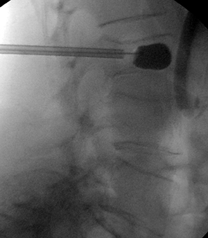
She underwent minimally invasive kyphoplasty to treat the fracture. Through a pen hole incision a balloon is placed inside the bone to restore the bone back to normal shape as possible. (It never looks quite normal). Then cement is placed into the bone and acts like grout making the fracture all healed.
Related Topics:
- Lumbar Laminectomy
- Posterior Lumbar Fusion
- Lumbar Endoscopic Discectomy
- Minimally Invasive Lumbar Discectomy
- Anterior Lumbar Interbody Fusion
- Minimally Invasive TLIF
- Kyphoplasty
- Minimally Invasive Spine Surgery
- Oblique Lumbar Interbody Fusion (OLIF)
- Posterior Cervical Laminectomy and Fusion
- Cervical Corpectomy and Strut Graft
- Endoscopic Spine Surgery
- Surgery for Scoliosis
- Cervical Laminoplasty
- Image-Guided Spine Surgery
- Anterior Cervical Discectomy with Fusion
- Artificial Cervical Disk Replacement
- Cervical Foraminotomy
- Extreme Lumbar Interbody Fusion