Radiculopathy/Myelopathy
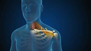
Cervical Spine Anatomy
The cervical spine is made up of 7 bones and in between each bone is a disc. The disc provides flexibility and motion to the spine. However, as we age, the disc can degenerate and lose some of its resiliency. The disc can have a tear in it and the material protrudes out or as the disc does not function normally, bone spurs can form because the motion is not as fluid as before.
What is Cervical Radiculopathy/Myelopathy?
Disk protrusion, also called herniated disk, is a condition caused by a tear in an intervertebral disk, allowing the disk contents to bulge out.
Disk protrusions in the cervical or neck area place pressure on nerve roots (nerve root compression) resulting in radiculopathy or on the spinal cord causing myelopathy.
Radiculopathy is a medical term used to describe the neurological deficits that can occur from pressure on the nerves which causes arm or finger weakness, numbness, and pain; this is the equivalent of sciatica in the arms. However, if the compression is of the spinal cord, the result is myelopathy which presents with hand clumsiness, burning pain the hands, and gait difficulty. If the compression becomes severe it can result in paralysis. Fortunately, both cervical radiculopathy and myelopathy can be treated if diagnosed promptly.
Causes of Cervical Radiculopathy/Myelopathy
Conditions that can cause radiculopathy/myelopathy include:
- Degenerative disk disease: Wear and tear of the disks between the vertebrae, causing them to lose their cushioning ability
- Spinal stenosis: Narrowing of the spinal canal as we age, most commonly due to degenerative arthritis
- Degenerative spondylolisthesis: Degeneration (wear and tear) of the vertebral components, usually occurring after age 50, causing slippage of a vertebra onto another, spinal stenosis and narrowing of the spinal canal
- Herniated Disc: Herniation of a disk is an anomalous spine condition characterized by the bulging of the inner contents of the intervertebral disk due to cracks in its outer wall.
Diagnosis of Cervical Radiculopathy/Myelopathy
In addition to a complete history and physical examination, your doctor may order spine X-ray, MRI or CT scans, electromyography and nerve conduction studies to diagnose cervical radiculopathy and myelopathy.
Treatment Options for Cervical Radiculopathy/Myelopathy
When conservative treatment measures such as rest, medication, physical therapy, and pain-blocking injections are ineffective, your surgeon may recommend spine surgery. Fortunately, radiculopathy can be managed nonoperatively in many cases and surgery is last resort. Unfortunately, myelopathy is a surgical problem that is only treated with surgery. It will get worse over time.
The most common spine surgery to relieve your symptoms of nerve root compression involves removing the disk and fusing the two vertebrae above and below it with a bone graft. This is called anterior cervical discectomy and fusion (ACDF). A newer treatment option is now available to replace the herniated disk with an artificial disk. Artificial disks are used in place of a bone fusion to preserve your neck’s movement and flexibility.
Sometimes however, if many discs are involved (more than 3), you may need a surgery from the back of your neck where a portion of the bone or lamina is removed. A spinal fusion with screws and rods are then inserted typically to stabilize the spine. This can cause loss of motion. To help preserve motion and flexibility, a different technique can utilized to reshape the shape of the bone without fusing your spine. This procedure is called a laminoplasty.
Case Study
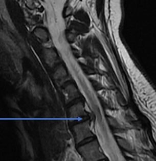
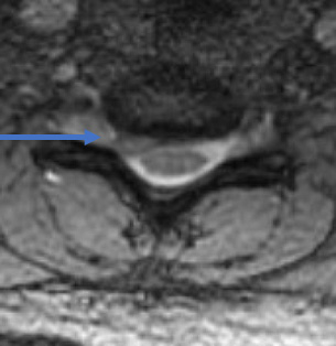
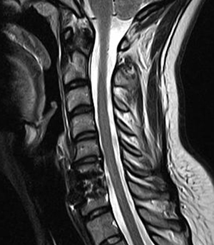
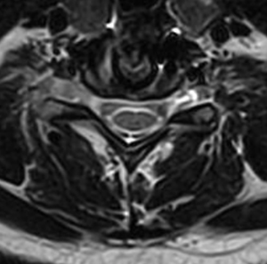
32 year old female comes with right arm pain that radiates all the way down to her fingers. We tried therapy, injections, oral pain medicine, and time. Because of the pain, a MRI showed a herniated disc in her neck on the right side (arrow). The left image is a side view and the right images is a cross section view.
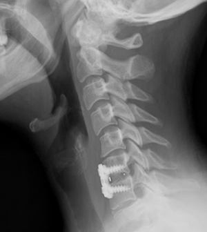
She underwent a minimally invasive anterior cervical discectomy and fusion after removing the herniated disc. A MRI shows resolution of the herniated disc.
- Cervical Corpectomy
- Posterior Cervical Fusion
- Cervical Laminoplasty
- Cervical Disk Replacement
- Cervical Discectomy With Fusion
- Case Study 3
- Case Study 5
- Case Study 8
- Case Study 9