Lumbar Discectomy
What is Lumbar Discectomy?
A lumbar discectomy is a surgical procedure performed to treat a herniated or ruptured disk and relieve pressure on the spinal nerves.
Lumbar Discectomy Procedure
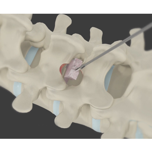
To perform lumbar discectomy, your doctor makes a small incision in your lower back over the affected spinal disk. Some vertebral bone and ligament may need to be removed to expose the disk. A microscope is used to visualize the disk and the adjacent spinal nerves. The spinal nerves are protected, and the affected disk is completely removed. The surgical site is then irrigated with antibiotic solution and closed. With new technologies, this procedure can be done more minimally invasively via endoscopic means.
Postoperative Care following Lumbar Discectomy
Following surgery, activities such as bending, lifting and sitting for prolonged periods should be avoided for four weeks. Your doctor will advise you about the exercises that will improve the strength and flexibility of the lower back. You may be able to return to work in 2-6 weeks depending on the level of activity involved.
Risks and Complications of Lumbar Discectomy
Lumbar discectomy, as with any invasive surgery may be associated with certain complications such as include nerve and spinal cord injury, infection and ongoing pain. Other risk include incomplete removal of the disc, reherniation, and leakage of spinal fluid. If bleeding continues after surgery, it can cause nerve compression that may need surgery again.
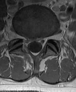
Clinical Example
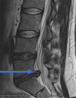
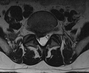
31 year old man presents with excruciating right leg pain. MRI shows a right L5-S1 disc herniation on the right (see arrow). The left image is a sagittal view where you are seeing the spine from the side. The right image is looking at a cross section of your spine. On this view the right side is the left of the image and vice versa.
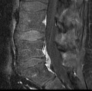
After minimally invasive surgery, the disc herniation is removed. The remaining “normal” disc is left alone.
Related Topics:
- Lumbar Laminectomy
- Posterior Lumbar Fusion
- Lumbar Endoscopic Discectomy
- Minimally Invasive Lumbar Discectomy
- Anterior Lumbar Interbody Fusion
- Minimally Invasive TLIF
- Kyphoplasty
- Minimally Invasive Spine Surgery
- Oblique Lumbar Interbody Fusion (OLIF)
- Posterior Cervical Laminectomy and Fusion
- Cervical Corpectomy and Strut Graft
- Endoscopic Spine Surgery
- Surgery for Scoliosis
- Cervical Laminoplasty
- Image-Guided Spine Surgery
- Anterior Cervical Discectomy with Fusion
- Artificial Cervical Disk Replacement
- Cervical Foraminotomy
- Extreme Lumbar Interbody Fusion